When Doctors Recommend DTI for Traumatic Brain Injuries
Your scan came back normal. But you still do not feel like yourself. That frustrating gap between how you feel and what standard imaging shows is something many traumatic brain injury patients experience. The brain can sustain real damage that conventional MRI simply cannot capture.
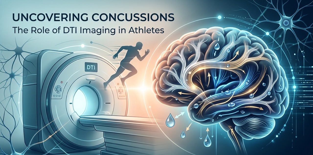
DTI for TBI changes that. Diffusion Tensor Imaging gives doctors a detailed look at white matter pathways inside the brain, revealing disruptions that explain persistent symptoms after head trauma. In this blog, you will learn when and why doctors recommend DTI, which conditions benefit most from it, and where to access advanced brain imaging in South Florida.
What Is DTI and Why Is It Used for Brain Injury Evaluation?
Diffusion Tensor Imaging is an advanced MRI technique that evaluates the white matter pathways running through the brain. These pathways are the brain’s internal communication network, connecting different regions and supporting thinking, movement, memory, and coordination.
Standard MRI shows brain structure. DTI goes further by measuring how water molecules move along nerve fibers. Disrupted movement patterns reveal damage to these pathways at a microscopic level.
For traumatic brain injury patients, this distinction matters. DTI detects injuries that conventional imaging misses and provides objective data that complements clinical assessments. It gives doctors a more complete picture when symptoms persist but standard scans appear normal.
Doctors May Recommend DTI When Symptoms Persist After a Head Injury
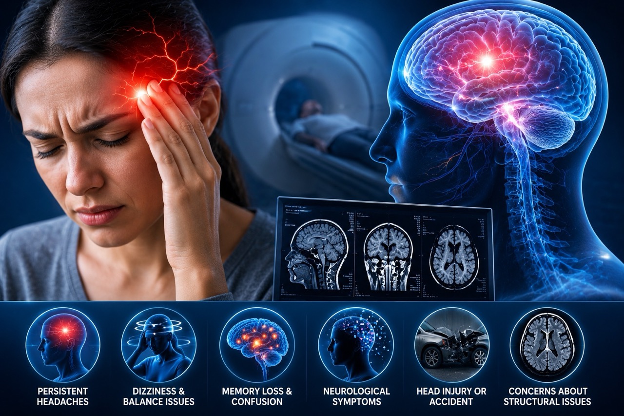
Some head injury symptoms linger far beyond what patients and doctors expect. When that happens, advanced imaging provides the next level of diagnostic detail. DTI may be considered when any of the following symptoms continue after a head injury without a clear explanation from standard scans:
Ongoing Headaches and Migraines
Headaches that return daily or worsen over time after a head injury may reflect damage to nerve fiber pathways involved in pain regulation. DTI can identify white matter disruptions in these regions. This information helps physicians understand the neurological basis of post-injury headaches and guides more targeted treatment rather than relying on symptom management alone.
Memory and Concentration Problems
Struggling to remember conversations, losing focus at work, or feeling mentally slower than usual are common complaints after traumatic brain injury. These symptoms often point to damage in the cognitive networks that support attention and memory formation. DTI evaluates the integrity of these specific pathways, giving clinicians measurable data to explain what patients are experiencing and why.
Dizziness and Balance Difficulties
Persistent dizziness and unsteadiness after a head injury suggest disruption in the neural circuits that manage spatial awareness and motor coordination. Standard imaging frequently misses this type of damage. DTI maps the pathways responsible for balance and identifies where connectivity has been compromised. This level of detail supports more accurate diagnosis and better-informed rehabilitation planning.
Brain Fog and Cognitive Changes
Brain fog is one of the most reported and least understood symptoms after traumatic brain injury. Patients describe it as mental heaviness, slow thinking, and difficulty processing information. DTI evaluates the white matter tracts that support cognitive processing speed and executive function. When these tracts show disruption, it confirms a neurological basis for what patients are experiencing beyond what standard scans reveal.
DTI May Be Recommended After a Concussion
Concussions fall under the umbrella of mild traumatic brain injury, but their effects are not always mild or short-lived. Many patients recover within a few weeks. Others continue experiencing symptoms for months. When recovery stalls without a clear structural explanation, physicians look for more detailed imaging options.
DTI provides additional information during this evaluation process by revealing microscopic damage to nerve fibers that conventional MRI cannot detect. It does not replace clinical assessments or neurological examinations. Rather, it adds an objective layer of data that helps doctors understand why symptoms persist and how to direct treatment more precisely for each patient’s specific injury profile.
Doctors Often Consider DTI for Post-Concussion Syndrome
Post-concussion syndrome develops when concussion symptoms extend well beyond the typical recovery window. It can significantly affect quality of life and daily function. When standard imaging does not explain the ongoing symptoms, DTI becomes a valuable next step in the evaluation process.
Physicians may consider DTI for post-concussion syndrome when patients report the following:
- Persistent Headaches. Daily or recurring headaches that do not respond to standard treatment may indicate ongoing white matter disruption in pain-processing regions of the brain.
- Difficulty Focusing. Patients who cannot sustain attention at work or school, or who feel mentally scattered without explanation, may have measurable damage to the neural pathways supporting cognitive focus.
- Mood and Behavioral Changes. Increased irritability, anxiety, or emotional instability after a concussion can reflect damage to the white matter circuits regulating emotional control and behavioral responses.
- Ongoing Cognitive Symptoms. Slower thinking, word-finding difficulties, and reduced mental stamina that persist long after injury may be visible on DTI even when conventional scans show no abnormality.
DTI May Be Used Following Motor Vehicle Accidents
Doctors may recommend DTI after a motor vehicle accident when patients experience ongoing neurological symptoms such as headaches, memory issues, dizziness, or concentration difficulties. Car accidents generate powerful forces that can injure the brain even without a direct blow to the head. Whiplash causes the brain to shift rapidly inside the skull, stretching and disrupting nerve fibers in ways that produce real neurological symptoms.
Many accident patients report headaches, cognitive changes, dizziness, and emotional shifts that begin hours or days after the collision. When these symptoms persist and standard imaging does not explain them, DTI for TBI offers the next level of diagnostic clarity. Advanced brain imaging in these cases supports both medical treatment and documentation needs for patients navigating recovery after a motor vehicle accident.
Athletes and Sports-Related Brain Injuries May Benefit From DTI Evaluation
Repeated head impacts in contact sports create cumulative risks that deserve serious neurological attention. Football, soccer, hockey, basketball, and combat sports athletes absorb collisions that can disrupt white matter pathways over time. A single concussion may resolve fully. Multiple concussions, or a concussion with prolonged symptoms, raises more complex questions about brain health.
Neurologists and sports medicine specialists sometimes recommend DTI when standard imaging does not explain persistent post-concussion symptoms in athletes. The imaging data helps guide return-to-play decisions and long-term brain health monitoring. Returning to activity before the brain has healed increases injury risk significantly and requires objective evidence to evaluate safely.
How DTI Supports a More Comprehensive Traumatic Brain Injury Assessment
Traumatic brain injury evaluation often involves multiple diagnostic tools working together. DTI is one component of a broader neurological assessment process. Here is how it fits alongside the other tools physicians use:
Neurological Examinations
A neurological examination assesses reflexes, coordination, cognition, and sensory function. It establishes a clinical baseline and identifies which areas of brain function may be affected. This examination gives physicians a framework for interpreting imaging findings in the context of the patient’s actual functional presentation.
Symptom Assessments
Structured symptom assessments document the type, severity, and duration of post-injury complaints. They help clinicians track changes over time and identify symptom clusters that point toward specific neurological concerns. This information guides imaging decisions and helps determine whether advanced techniques like DTI are warranted based on the patient’s evolving clinical picture.
Traditional MRI Studies
Conventional MRI identifies structural abnormalities including bleeding, swelling, and lesions. It provides an important first layer of evaluation after traumatic brain injury. However, its limitations in detecting microscopic white matter damage mean it does not always explain why patients continue experiencing symptoms. This gap is where DTI adds measurable value to the diagnostic process.
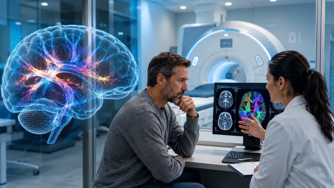
Advanced DTI Imaging
DTI evaluates white matter integrity and brain connectivity at a level no other standard imaging technique reaches. It detects disruptions in nerve fiber pathways caused by traumatic brain injury and maps where these disruptions occur. Combined with clinical findings and conventional MRI, DTI gives physicians the most complete picture available for evaluating TBI and guiding individualized treatment decisions.
Where to Access Advanced DTI Imaging in South Florida
Choosing the right imaging center for DTI evaluation matters. Advanced neuroimaging requires experienced radiologists, current technology, and a team that understands the clinical context of brain injury.
Precision MRI Group offers DTI brain scans at our Cypress Creek location following a recent equipment upgrade that expanded our advanced neuroimaging capabilities. Our board-certified radiologists review every scan with the detail that complex cases demand.
We deliver reports within 24 to 48 hours. Same-day appointments are available for patients who need timely evaluation. Late evening and weekend scheduling accommodates patients with demanding schedules. Free transportation is provided on request. Our multilingual staff speaks English, Spanish, and Creole to support clear communication for every patient we serve.
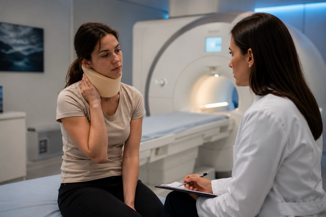
When Should You Discuss DTI With Your Doctor?
If you have experienced a head injury and your symptoms have not resolved, it may be time to ask your doctor about advanced imaging options.
Bring up DTI if you are dealing with any of the following after a traumatic brain injury:
- Chronic Headaches. Headaches that persist weeks or months after your injury without improvement despite treatment deserve further neurological investigation.
- Memory Difficulties. Trouble recalling information, retaining new details, or maintaining mental clarity after a head injury may reflect white matter damage that DTI can detect.
- Dizziness. Ongoing dizziness or unsteadiness that affects daily life suggests disruption in the neural circuits managing balance and spatial orientation.
- Cognitive Impairment. Slower processing speed, reduced decision-making ability, and difficulty with complex tasks are neurological symptoms that warrant advanced imaging evaluation.
- Balance Issues. Persistent problems with coordination or gait after a head injury may indicate damage in motor control pathways that DTI can specifically evaluate.
- Prolonged Concussion Symptoms. When expected recovery timelines pass and symptoms remain, DTI provides the diagnostic depth needed to understand why and what to do next.
Get the Brain Injury Answers You Deserve
Persistent symptoms after a traumatic brain injury are not something to push through or dismiss. Physicians recommend DTI for TBI when standard scans fall short, symptoms persist, or recovery takes longer than expected. It provides objective neurological data that supports better diagnosis, smarter treatment planning, and more informed decisions about returning to normal activity.
Early evaluation leads to better outcomes. Do not wait for symptoms to worsen before seeking comprehensive care. Contact Precision MRI Group today to learn more about advanced MRI and DTI imaging options available across South Florida. Same-day appointments are available at our Cypress Creek location and beyond.
Precision MRI Group Locations:
Cypress Creek (DTI Available)
2122 NW 62nd Street, Suite 107, Ft. Lauderdale, FL 33309
Phone: (954) 677-1069, Contact: Latoya Reid (latoya@cypresscreekmri.com)
Additional Locations:
Pembroke Pines
9696 Pines Blvd., Pembroke Pines, FL 33024
Phone: (954) 391-7844, Contact: Amalia (amalia@pinesimagingcenter.com)
Lake Worth
2311 10th Ave N Suite #2 and Suite #1, Lake Worth, FL 33461
Phone: (561) 623-8346, Contact: Marisol (marisol@mriprecision.com)
Port St Lucie
879 E Prima Vista Blvd #2, Port St. Lucie, FL 34952
Phone: (772) 344-7566, Contact: Laura Schwenzer (laura@mriprecision.com)