MRI Brain Tumor Detection: How Scans Reveal Hidden Problems
Experiencing persistent headaches, vision changes, or unexplained symptoms creates worry about serious health conditions. Many people wonder if their symptoms indicate a brain tumor requiring immediate medical attention. Getting accurate answers quickly helps reduce anxiety and guides appropriate treatment when needed most.
An MRI brain tumor scan is the most effective imaging test for detecting abnormal growths. This advanced technology reveals soft tissue details that other tests cannot show clearly or accurately. Furthermore, MRI brain tumor detection accuracy exceeds 90% for identifying masses requiring medical evaluation.
At Precision MRI Group, we understand that waiting for answers creates stress and uncertainty daily. Our facilities offer fast scheduling with appointments available within days rather than weeks or months. Board-certified radiologists deliver detailed reports within 24 to 48 hours for quick diagnosis decisions. Additionally, our experienced team makes the entire process comfortable and stress-free throughout your visit.
How MRI Detects Brain Tumors
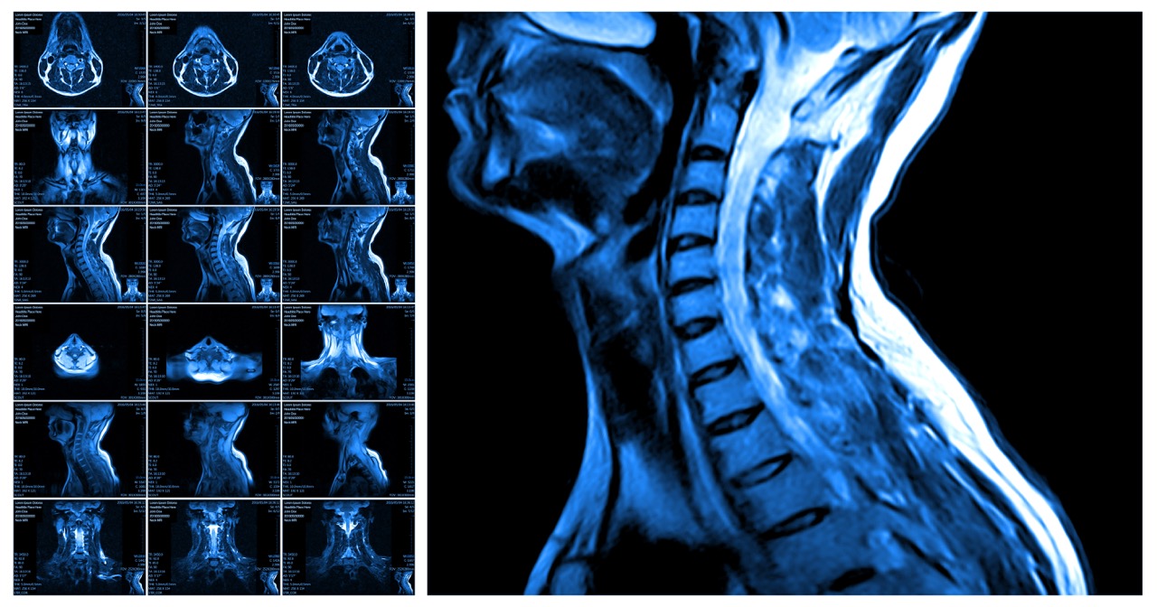
MRI brain tumor imaging uses powerful magnetic fields and radio waves to create detailed pictures. The technology shows soft tissues, blood vessels, and abnormal growths with exceptional clarity and precision. Unlike CT scans that primarily show bone, MRI reveals brain tissue changes indicating tumor presence.
MRI brain tumor detection is preferred because it doesn’t use radiation and provides superior detail. Radiologists examine images looking for masses, swelling, pressure on surrounding tissues, and abnormal changes. These indicators help distinguish between tumors, cysts, inflammation, and other conditions requiring different treatments.
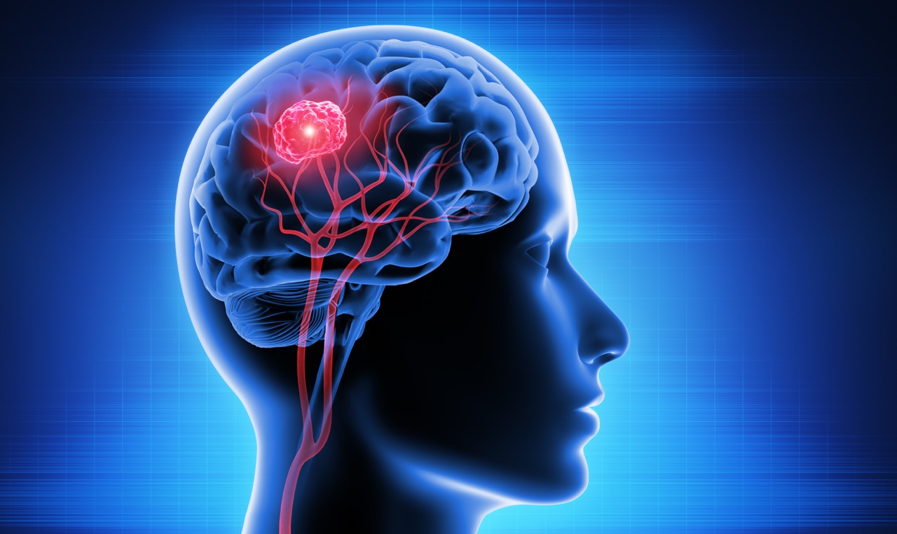
The MRI brain tumor scan reveals several critical features that guide diagnosis and treatment:
- Tumor size – Measurements help determine growth rate and treatment urgency for patient safety.
- Location – Position affects which brain functions might be impacted and surgical approach planning.
- Swelling – Surrounding edema indicates how the tumor affects nearby healthy brain tissue significantly.
- Tissue enhancement (with contrast) – Shows blood vessel patterns that help classify tumor type accurately.
MRI With and Without Contrast for Brain Tumor Detection
MRI brain tumor scans can be performed with or without contrast dye injection. Without contrast, MRI shows structural changes, mass size, and tissue density differences clearly. This basic scan often provides enough information for initial evaluation and screening purposes effectively.
With contrast, gadolinium dye highlights tumor borders, blood vessels, and inflammation with greater detail. The enhanced images show how tumors take up contrast differently than normal tissue. Furthermore, MRI brain tumor imaging with contrast helps doctors distinguish between tumor types and treatment responses.
When Contrast Is Recommended
Doctors order contrast-enhanced MRI brain tumor scans when they suspect abnormal growths need evaluation. Monitoring tumor growth over time requires contrast to track size changes and treatment effects. Tracking treatment progress after surgery, radiation, or chemotherapy needs contrast enhancement for accuracy.
Contrast helps radiologists see small tumors that might be invisible on non-contrast scans alone. The enhanced images reveal blood vessel patterns unique to different tumor types requiring specific treatments. Additionally, post-surgical scans use contrast to distinguish between scar tissue and tumor recurrence clearly.
When Non-Contrast MRI Is Sufficient
Initial evaluation of neurological symptoms often starts with non-contrast MRI brain tumor screening. Doctors order these scans to rule out obvious masses before ordering more detailed studies. Patients who cannot receive contrast due to kidney problems or allergies get non-contrast scans.
Follow-up imaging for stable, benign tumors may not need contrast at every appointment. Non-contrast scans work well for evaluating brain structure and detecting large abnormalities effectively.
Symptoms That May Require a Brain MRI
Certain symptoms warrant brain imaging to rule out tumors or other serious conditions. Early detection through MRI brain tumor screening improves treatment outcomes significantly when problems exist. Never ignore persistent symptoms that disrupt your daily life or worsen over time.
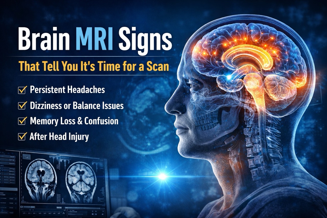
Here are symptoms that may indicate you need a brain MRI evaluation:
- Persistent headaches – New, severe, or worsening headaches, especially with morning vomiting, need evaluation promptly.
- Vision changes – Blurred vision, double vision, or peripheral vision loss may indicate pressure on nerves.
- Seizures – New-onset seizures in adults without prior history warrant immediate imaging for diagnosis always.
- Balance issues – Unexplained dizziness, coordination problems, or falls suggest possible neurological problems requiring assessment.
- Nausea and vomiting – Persistent symptoms without clear cause, especially morning sickness, may indicate pressure buildup.
- Memory or personality changes – Confusion, mood swings, or behavioral changes can signal brain tissue problems.
- Weakness or numbness – Progressive weakness or numbness in limbs suggests possible nerve compression needing evaluation.
Can an MRI Miss a Brain Tumor?
MRI brain tumor detection is highly sensitive, identifying most masses requiring medical treatment effectively. However, extremely small lesions under 2-3 millimeters might not appear on standard imaging protocols. Rare cases involve tumors in difficult locations that blend with normal tissue on scans.
High-resolution scanners like 1.5 Tesla and 3 Tesla technology improve detection accuracy significantly for small lesions. These advanced machines capture more detailed images than older, lower-strength scanners some facilities use. Furthermore, experienced radiologists trained in neuroimaging recognize subtle changes others might miss completely. Precision MRI Group uses 1.5 Tesla technology that provides excellent detail for brain tumor detection. Our board-certified radiologists specialize in interpreting brain imaging with expertise and precision consistently.
How Radiologists Interpret a Brain MRI
Reading MRI brain tumor scans requires specialized training and experience in neuroimaging interpretation consistently. Radiologists examine tissue density differences that indicate abnormal cell growth or masses present. Enhancement patterns after contrast injection reveal how tumors receive blood supply and behave biologically.
Swelling and pressure on surrounding brain structures indicate how aggressive tumors are currently. Blood flow patterns help distinguish between tumor types requiring different treatment approaches and protocols. Structural shifts show if tumors are pushing brain tissue aside or invading surrounding areas.
Board-certified radiologists at Precision MRI Group provide detailed reports describing all findings accurately. Their expertise ensures subtle abnormalities don’t get missed during image interpretation and analysis processes. Furthermore, timely reports within 24 to 48 hours help doctors plan treatment quickly.
Brain Conditions an MRI Can Identify
MRI brain tumor imaging also detects many other conditions affecting brain health and function. The comprehensive evaluation provides doctors with information about various neurological problems requiring treatment. A single scan can reveal multiple issues that explain your symptoms completely and accurately.
Here are conditions MRI identifies beyond tumors:
- Benign tumors – Non-cancerous growths like meningiomas that may need monitoring or removal depending on location.
- Malignant tumors – Cancerous growths requiring immediate treatment with surgery, radiation, or chemotherapy for survival.
- Cysts – Fluid-filled sacs that may cause pressure symptoms but often don’t require treatment unless large.
- Abscesses – Infections that create pus-filled areas needing antibiotic treatment or surgical drainage for resolution.
- Stroke – Blood vessel blockages or bleeding that cause brain tissue damage requiring emergency medical intervention.
- Multiple sclerosis lesions – Areas of inflammation that indicate autoimmune disease affecting nerve function significantly.
- Inflammation – Swelling from infections, autoimmune conditions, or other causes affecting brain tissue health overall.
Brain MRI diagnosis capabilities extend far beyond tumor detection for comprehensive neurological evaluation. MRI findings brain scans reveal guide appropriate treatment for numerous conditions affecting your health.
Why Choose Precision MRI Group for Brain MRI
Precision MRI Group provides comprehensive MRI brain tumor imaging with advanced technology and expert interpretation. Our facilities throughout Florida offer convenient access to high-quality neuroimaging services for everyone. We understand that brain symptoms create anxiety and need prompt evaluation for peace of mind.
Fast appointments mean you get scanned within days when symptoms require urgent evaluation and assessment. Our 24 to 48-hour reports ensure your doctor receives results quickly for treatment planning decisions. Multilingual staff speaking English, Spanish, and Creole ensures clear communication with all patients consistently.
Short-bore MRI design reduces claustrophobia and anxiety for patients nervous about traditional closed machines. Free transportation upon request removes barriers for patients who have difficulty getting to appointments. Our 1.5 Tesla high-resolution imaging provides exceptional detail for accurate MRI brain tumor detection consistently.
Brain MRI Pembroke Pines, Lake Worth, Cypress Creek, and Port St. Lucie locations serve patients across South Florida. Brain MRI Florida services at Precision MRI Group combine technology with compassionate care always.
Preparing for Your Brain MRI
Simple preparation ensures your MRI brain tumor scan produces the clearest possible images for diagnosis. Taking a few minutes to prepare properly prevents delays and ensures optimal scan quality. Most patients find the preparation straightforward and easy to follow without complications occurring.
Follow these practical steps for the best scanning experience:
- Remove metal – Take off jewelry, watches, glasses, hearing aids, and all metal accessories before scanning.
- Bring medical history – Provide information about symptoms, medications, and previous imaging or surgeries relevant.
- Stay still – Remaining motionless during scanning ensures clear images without blurriness affecting diagnostic quality.
- Discuss claustrophobia – Inform staff if you feel anxious in enclosed spaces for accommodation options.
- Understand contrast instructions – Ask questions about contrast dye if ordered for your specific scan.
Prep for brain MRI takes just a few minutes when you arrive prepared and informed. What to expect MRI information helps reduce anxiety about the unknown scanning experience ahead.
Book Your Brain MRI at Precision MRI Group
Don’t let worrying symptoms go undiagnosed when answers are available quickly at our facilities. Schedule your MRI brain tumor scan at any of our four convenient Florida locations today. Our experienced team makes getting quality neuroimaging simple, comfortable, and stress-free for everyone always.
Same-day and next-day appointments accommodate urgent evaluation needs when symptoms cause concern or worry. Contact us today to schedule your scan and get the peace of mind you deserve.
Precision MRI Group Locations:
Pembroke Pines
9696 Pines Blvd., Pembroke Pines, FL 33024
Phone: (954) 391-7844, Contact: Amalia (amalia@pinesimagingcenter.com)
Lake Worth
2311 10th Ave N Suite #2 and Suite #1, Lake Worth, FL 33461
Phone: (561) 623-8346, Contact: Marisol (marisol@mriprecision.com)
Cypress Creek
2122 NW 62nd Street, Suite 107, Ft. Lauderdale, FL 33309
Phone: (954) 677-1069, Contact: Latoya Reid (latoya@cypresscreekmri.com)
Port St Lucie
879 E Prima Vista Blvd #2, Port St. Lucie, FL 34952
Phone: (772) 344-7566, Contact: Laura Schwenzer (laura@mriprecision.com)