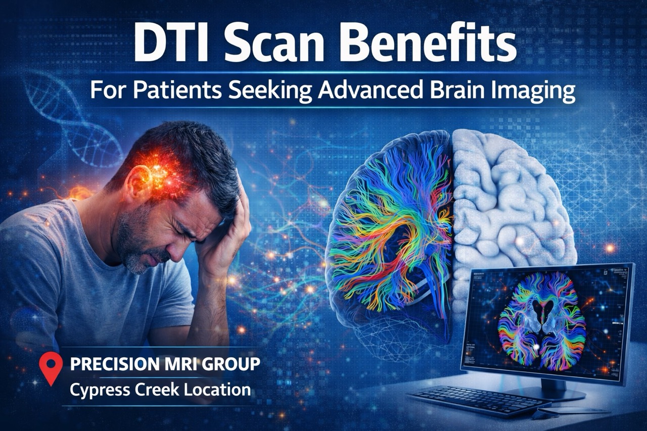
DTI Scan Benefits for Patients Seeking Advanced Brain Imaging
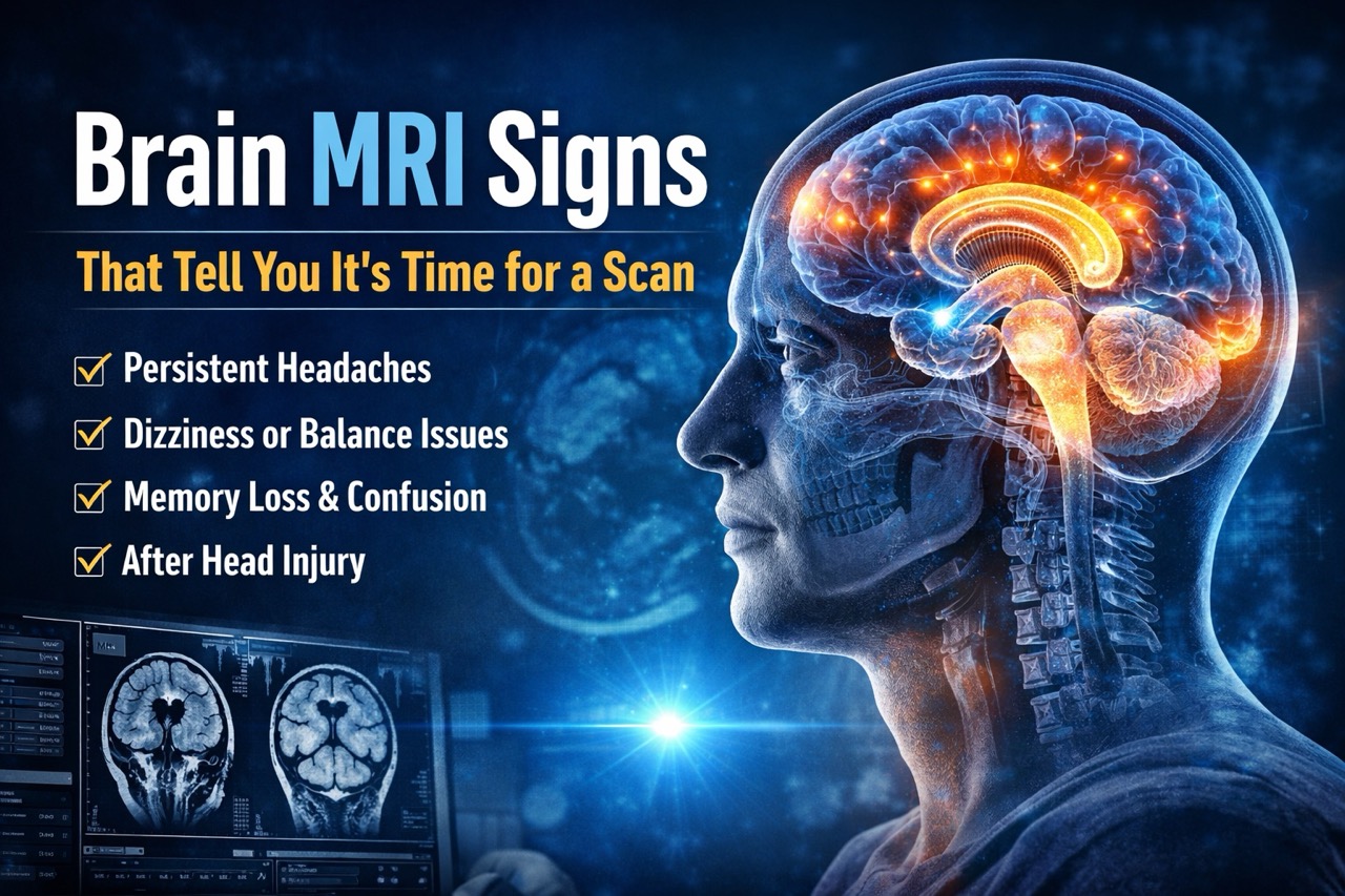
You hit your head weeks ago, but headaches and confusion still plague your daily life. Standard MRI came back normal, yet symptoms persist and affect your work and relationships. Your doctor mentions something called a DTI scan that might reveal what standard imaging missed.
Many patients feel frustrated when traditional tests show nothing despite real, debilitating symptoms continuing. Brain injuries don’t always appear on standard MRI, leaving patients without answers or proper treatment. Advanced imaging technology now detects subtle brain damage that older methods cannot see at all.
Precision MRI Group offers Diffusion Tensor Imaging at our Cypress Creek location for patients. This advanced neuroimaging technique reveals brain injuries and neurological conditions invisible on standard scans. Furthermore, DTI scan technology provides answers when other imaging leaves questions unanswered about your health.
Understanding What a DTI Scan Is
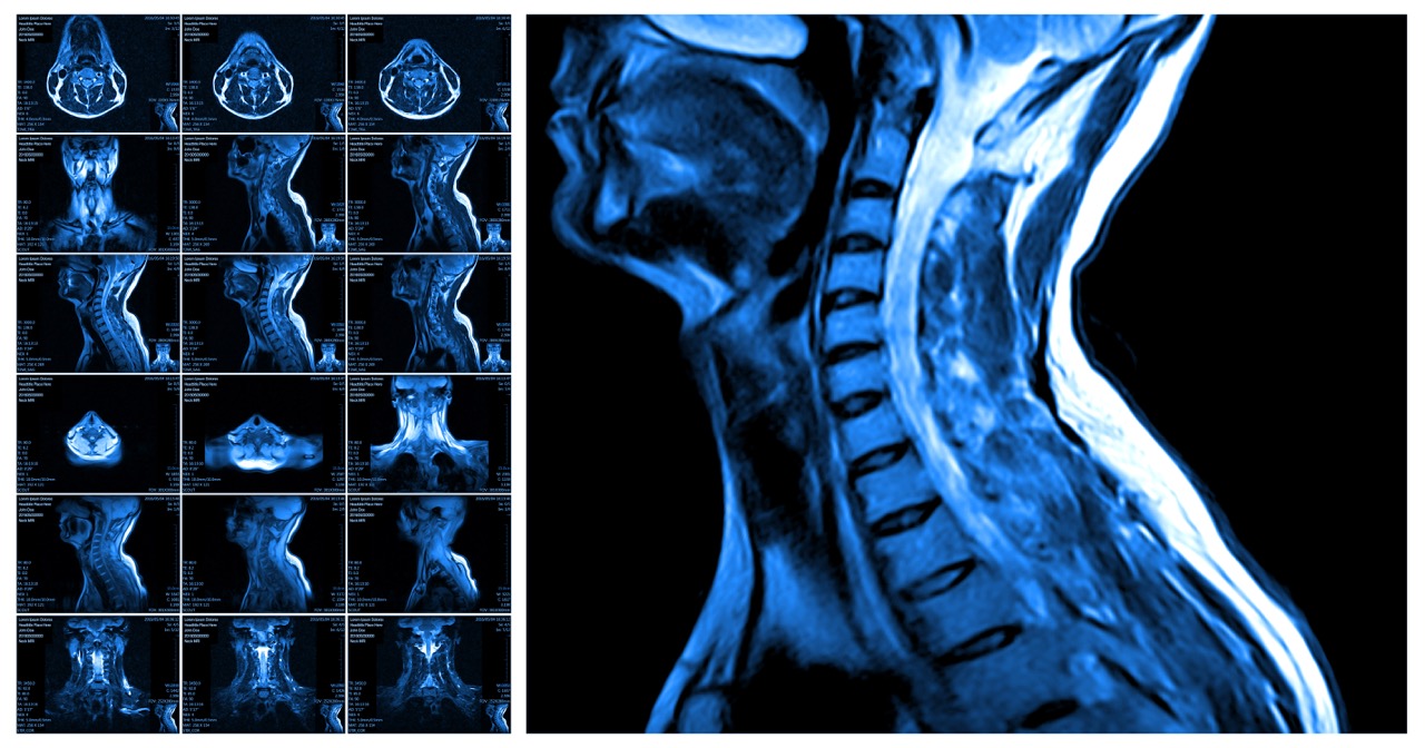
Diffusion Tensor Imaging, or DTI, is an advanced type of MRI that shows brain structure. The technology maps white matter pathways connecting different brain regions with remarkable precision and detail. These neural pathways carry signals between brain areas, controlling everything from movement to memory.
DTI scan technology tracks how water molecules move along nerve fibers in brain tissue. Damaged or disrupted pathways show abnormal water movement patterns radiologists can identify clearly. This reveals injuries to nerve fibers that don’t appear on conventional MRI images at all.
DTI is considered advanced neuroimaging because it shows microstructural changes standard MRI cannot detect. The technique provides information about brain connectivity and white matter integrity for diagnosis. Furthermore, DTI scan results help doctors understand injury severity and predict recovery potential accurately.
How a DTI Scan Works
DTI technology uses the same MRI machine as standard brain imaging but with specialized sequences. The scan measures how water molecules diffuse through brain tissue in multiple directions carefully. This diffusion data creates detailed maps showing the brain’s white matter structure and integrity.
Tracking Water Molecule Movement in Brain Tissue
Water naturally moves along nerve fibers in healthy brain tissue following predictable patterns consistently. Injured or damaged nerve pathways disrupt normal water movement, creating abnormal diffusion patterns clearly. DTI scan sequences measure this movement in at least six directions to map pathways. The technology calculates how freely water moves and in which directions throughout brain regions.
Identifying Microstructural Changes in White Matter
White matter contains nerve fibers connecting different brain areas for communication and function coordination. DTI reveals damage to these fibers even when brain structure appears normal on imaging. The scan shows areas where nerve pathways are disrupted, torn, or degenerating over time. Color-coded maps display healthy pathways versus damaged regions for visual assessment and comparison.
How Radiologists Interpret DTI Results
Radiologists analyze DTI images looking for areas with abnormal water diffusion patterns indicating damage. They compare your scan to normal brain patterns to identify significant differences requiring attention. Measurements like fractional anisotropy show white matter integrity quantitatively for objective assessment. Furthermore, experienced radiologists correlate DTI findings with your symptoms for accurate clinical diagnosis.
What Conditions Can a DTI Scan Detect?
DTI scan technology excels at identifying brain conditions that standard imaging often misses completely. Traumatic brain injuries and concussions damage nerve fibers without causing visible structural changes. Mild TBI symptoms like headaches, concentration problems, and mood changes finally have visible evidence.
Stroke-related white matter damage shows up clearly on DTI even when standard MRI looks normal. Neurodegenerative diseases like Alzheimer’s and Parkinson’s cause progressive white matter deterioration DTI reveals early. Complex neurological symptoms without clear cause often result from subtle brain pathway disruption.
Brain disorders involving disrupted neural pathways include multiple sclerosis, autism, and schizophrenia presentations. DTI helps doctors understand how these conditions affect brain connectivity and function over time. Furthermore, the technology provides objective evidence of injury for medical documentation and treatment planning.
DTI vs Standard MRI: Understanding the Difference
Both DTI and standard MRI use magnetic resonance technology but reveal different information. Understanding when each imaging type works best helps doctors order appropriate tests initially.
What Standard MRI Shows
Standard MRI excels at showing brain structure, tumors, bleeding, and large-scale tissue damage. The imaging reveals structural abnormalities like masses, fluid collections, and obvious tissue injuries. Doctors use standard MRI for most brain imaging needs because it’s widely available. However, standard MRI cannot detect microscopic damage to individual nerve fiber pathways reliably.
Why DTI Reveals Subtle Injuries MRI May Miss
DTI scan technology detects microstructural changes in white matter that don’t alter brain appearance. The imaging shows functional damage to nerve pathways even when tissue looks structurally normal. Concussions and mild traumatic brain injuries often damage connections without visible tissue destruction. Furthermore, DTI provides evidence of injury when patients have symptoms but normal standard imaging.
When Doctors Recommend One or Both Imaging Types
Standard MRI remains the first imaging test for most brain symptoms and concerns initially. DTI scan becomes necessary when standard imaging is normal but symptoms persist or worsen. Doctors order both when they need comprehensive evaluation of structure and white matter integrity. Furthermore, combining both imaging types provides the most complete picture of brain health.
Advanced DTI Brain Imaging Available at Precision MRI Group
Precision MRI Group now offers cutting-edge DTI scan technology at our Cypress Creek location. Our recent upgrade enables this advanced neuroimaging capability for patients needing detailed brain evaluation. This enhancement demonstrates our commitment to providing the most advanced diagnostic tools available today.
The upgraded Cypress Creek MRI machine supports DTI with the latest imaging protocols and sequences. This capability matters for patients experiencing persistent neurological symptoms without clear diagnosis on imaging. Enhanced diagnostic accuracy helps neurologists, concussion specialists, and personal injury physicians provide better care.
Physicians throughout South Florida can now refer patients for DTI when standard imaging proves insufficient. The technology supports comprehensive evaluation of traumatic brain injury, stroke, and neurodegenerative conditions. Personal injury attorneys value DTI’s ability to document invisible brain injuries for legal cases. Furthermore, this advanced neuroimaging serves concussion specialists treating athletes and accident victims with care.
When Patients Should Consider a DTI Scan
Certain symptoms and situations warrant advanced brain imaging beyond standard MRI scanning protocols available. DTI scan technology provides answers when conventional imaging leaves doctors and patients without explanations.
Consider DTI imaging if you experience these concerning symptoms or situations:
- Persistent symptoms after a concussion – Headaches, dizziness, or cognitive problems lasting weeks after injury.
- Memory changes, headaches, dizziness, mood issues – Neurological symptoms without clear cause on standard tests.
- Post-stroke changes that require more clarity – Understanding white matter damage extent for rehabilitation planning.
- Unexplained neurological symptoms – Balance problems, weakness, or sensory changes without identified cause yet.
- When physicians need higher diagnostic accuracy – Cases where treatment decisions depend on understanding injury severity.
What to Expect During a DTI Scan at Precision MRI Group
Getting a DTI scan at Precision MRI Group follows the same comfortable process as standard MRI. The experience is quick, painless, and requires no special preparation beyond standard MRI protocols. Skilled technologists oversee your scan and ensure optimal image quality throughout the procedure carefully.
Our Cypress Creek facility provides a welcoming environment with short-bore MRI for patient comfort. Multilingual staff speaking English, Spanish, and Creole ensures clear communication with all patients consistently. Free transportation to appointments removes barriers for patients who have difficulty getting to facilities.
Late-night and weekend scheduling accommodates busy work schedules and urgent evaluation needs without delay. The DTI scan itself takes about 45 to 60 minutes depending on sequences needed. Furthermore, our experienced team makes your advanced neuroimaging experience as comfortable as possible always.
How Fast Will DTI Scan Results Be Ready?
Precision MRI Group maintains its commitment to fast results even for advanced neuroimaging like DTI. Our standard 24 to 48-hour report turnaround applies to DTI scans just like other imaging. This quick turnaround helps doctors start treatment planning without unnecessary delays that worsen outcomes.
Immediate results are available upon request for urgent cases requiring rapid diagnosis and intervention. Emergency situations and time-sensitive medical decisions receive priority interpretation from our radiologists immediately. Board-certified radiologists specializing in neuroimaging review all DTI scans with expertise and precision.
Our radiologists understand DTI technology and how to interpret complex white matter findings accurately. Furthermore, detailed reports explain findings in language referring physicians can use for treatment decisions.
How DTI Scans Improve Patient Care
Advanced neuroimaging with DTI scan technology transforms how doctors diagnose and treat brain injuries. Better treatment planning results from understanding the true extent and location of brain damage. Early detection of invisible injuries prevents complications and supports appropriate intervention timing for recovery.
More accurate medical documentation helps patients receive proper treatment and disability benefits when needed. The objective evidence DTI provides supports workers’ compensation and personal injury claims with facts. Increased clarity for neurologists helps them understand complex cases that previously remained diagnostic mysteries.
Primary care doctors gain confidence referring patients when DTI confirms suspicions about brain injury presence. Attorneys representing injured clients value DTI’s ability to demonstrate invisible injuries with visual evidence. Furthermore, patients feel validated when imaging finally shows what they’ve been experiencing all along.
Get Answers With Advanced DTI Brain Imaging
DTI scan technology represents a major advancement in how we diagnose and understand brain injuries. This cutting-edge neuroimaging reveals damage that standard tests miss, providing answers patients desperately need. Precision MRI Group’s investment in DTI demonstrates our commitment to offering the most advanced care.
Our Cypress Creek location now provides this sophisticated brain imaging for patients throughout South Florida. Experience the difference that advanced technology and expert interpretation make for neurological diagnosis today.
Schedule your DTI scan today at Precision MRI Group’s Cypress Creek location and experience advanced neuroimaging designed for accuracy, comfort, and fast results.
Precision MRI Group Locations:
Cypress Creek (DTI Available)
2122 NW 62nd Street, Suite 107, Ft. Lauderdale, FL 33309
Phone: (954) 677-1069, Contact: Latoya Reid (latoya@cypresscreekmri.com)
Additional Locations:
Pembroke Pines
9696 Pines Blvd., Pembroke Pines, FL 33024
Phone: (954) 391-7844, Contact: Amalia (amalia@pinesimagingcenter.com)
Lake Worth
2311 10th Ave N Suite #2 and Suite #1, Lake Worth, FL 33461
Phone: (561) 623-8346, Contact: Marisol (marisol@mriprecision.com)
Port St Lucie
879 E Prima Vista Blvd #2, Port St. Lucie, FL 34952
Phone: (772) 344-7566, Contact: Laura Schwenzer (laura@mriprecision.com)