Understanding MRI Nerve Damage Results and What They Reveal
Nerve pain creates frustrating symptoms that disrupt your daily life and activities significantly every day. Shooting pain, tingling, numbness, and weakness make simple tasks feel impossible to complete comfortably. Many patients wonder if MRI can reveal what’s causing their nerve symptoms and discomfort.
Understanding MRI nerve damage detection helps you know what to expect from diagnostic imaging. Doctors often order MRI first because it shows structures surrounding nerves without invasive procedures. The technology reveals compression, inflammation, and abnormalities that cause nerve pain and dysfunction clearly.
At Precision MRI Group, we provide fast answers with advanced 1.5 Tesla imaging technology. Our experienced radiologists specialize in identifying nerve-related problems with expertise and precision consistently. Same-day appointments mean you don’t wait weeks while symptoms worsen or become unbearable daily. Furthermore, our comfortable facilities throughout Florida make getting quality nerve imaging simple and stress-free.
Can an MRI Show Nerve Damage?
MRI nerve damage detection works differently than most people expect from diagnostic imaging initially. The technology doesn’t always “see” individual nerves directly like it shows bones or organs. Instead, MRI reveals nerve compression from bulging discs, swelling around nerve pathways, and inflammation.
MRI nerve damage evaluation shows structural problems causing nerve symptoms with exceptional detail and clarity. Disc pressure on nerve roots appears clearly on detailed imaging sequences capturing multiple angles. Structural problems like bone spurs, tumors, or cysts pressing on nerves show up well.
The scan displays swelling in nerve pathways that indicates irritation or injury affecting function. Inflammation surrounding compressed nerves creates signal changes radiologists recognize as abnormal immediately. Furthermore, MRI nerve damage assessment guides treatment by showing exactly where problems occur.
What Nerve Problems MRI Can Detect
MRI technology excels at identifying structural causes of nerve pain and dysfunction throughout the body. Understanding what MRI nerve damage imaging reveals helps set realistic expectations for your scan. The detailed pictures show compression points, inflammation, and masses affecting nerve health significantly.
Here are nerve problems MRI commonly detects during diagnostic evaluation:
- Pinched nerve from disc bulge – Discs pressing on nerve roots create pain, numbness, and weakness.
- Herniated disc compressing nerves – Ruptured discs squeeze nerves causing severe radiating pain and symptoms.
- Sciatica and nerve root irritation – Lower spine nerve compression causing leg pain and dysfunction.
- Spinal stenosis – Narrowed spinal canal squeezing nerves and spinal cord progressively over time.
- Nerve inflammation – Swelling around nerves from injury, infection, or autoimmune conditions clearly visible.
- Tumors affecting nerves – Masses pressing on or invading nerve tissue causing progressive symptoms.
- Post-injury nerve swelling – Trauma-related inflammation and compression from bleeding or edema accumulation.
When You Should Get an MRI for Nerve Pain
Timing matters when deciding whether you need MRI nerve damage evaluation for your symptoms. Severe pain that limits daily activities or prevents sleep warrants imaging sooner rather than later. Numbness or weakness in arms or legs suggests nerve compression requiring prompt evaluation always.
Pain lasting over four to six weeks without improvement indicates structural problems needing diagnosis. Radiating pain that shoots down arms or legs follows nerve pathways requiring imaging assessment. Spine injuries from accidents or falls often cause nerve damage needing MRI confirmation quickly.
Progressive symptoms that worsen over time suggest ongoing nerve compression requiring intervention soon. Loss of bladder or bowel control requires emergency evaluation including immediate MRI scanning. Furthermore, MRI nerve damage detection helps doctors plan appropriate treatment before permanent damage occurs.
MRI vs EMG: Which Test Shows Nerve Damage Better?
Different tests evaluate different aspects of nerve problems for comprehensive diagnosis and understanding. MRI nerve damage imaging shows structural causes while EMG measures nerve function electrically. Combining both tests often provides the most complete picture of nerve health overall.
What MRI Shows (Structural Issues)
MRI reveals discs pressing on nerves, joint problems causing compression, and soft tissue swelling. The imaging shows tumors affecting nerve pathways and spinal alignment issues clearly throughout. Structural abnormalities like bone spurs, cysts, or inflammation appear on MRI sequences distinctly.
What EMG Shows (Nerve Function)
EMG tests measure electrical activity traveling through nerves to muscles during contraction and rest. Nerve conduction studies show how fast signals travel along nerve pathways precisely measured. Muscle response testing reveals whether nerves communicate effectively with muscles they control daily. Furthermore, MRI nerve damage imaging and EMG complement each other for complete evaluation.
Types of MRI Used for Nerve Evaluation
Different MRI types target specific nerve problems depending on symptom location and patterns. Your doctor orders the scan type most likely to reveal your nerve damage. MRI nerve damage protocols vary based on which nerves need evaluation for accurate diagnosis.
Here are common MRI types used for nerve evaluation:
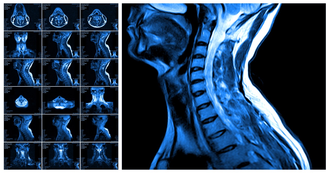
- Spine MRI – Evaluates cervical, thoracic, or lumbar nerve roots compressed by discs or stenosis.
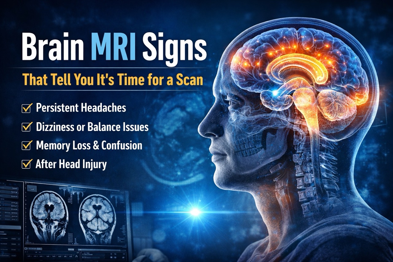
- Brain MRI (for facial nerve, trigeminal nerve, central causes) – Shows cranial nerves and brain conditions causing symptoms.
- Extremity MRI – Examines peripheral nerves in arms, legs, hands, or feet for compression points.
- MR Neurography (if relevant) – Specialized sequences highlight peripheral nerves with enhanced detail when available.
What to Expect During Your MRI at Precision MRI Group
Getting MRI nerve damage imaging at our facilities is comfortable and straightforward for all patients. Our short-bore MRI scanners provide more open space than traditional closed machines significantly reducing anxiety. Headphones with music make the experience pleasant while blocking loud scanner sounds effectively.
Experienced technologists guide you through positioning and explain what to expect during scanning procedures. Same-day scheduling means you get answers quickly when nerve pain needs urgent evaluation. Free transportation upon request removes barriers for patients who have difficulty getting to appointments.
Multilingual staff speaking English, Spanish, and Creole ensures clear communication with everyone consistently. The scan typically takes 30 to 45 minutes depending on body areas examined thoroughly. Furthermore, our comfortable environment helps patients relax during imaging for the best picture quality.
How Precision MRI Group Helps Diagnose Nerve Pain Faster
Fast diagnosis helps you start treatment before nerve damage becomes permanent or irreversible over time. Our board-certified radiologists deliver reports within 24 to 48 hours for quick treatment planning. These specialists have extensive experience identifying MRI nerve damage patterns and subtle findings others miss.
Late-night and weekend appointments accommodate busy schedules and urgent evaluation needs without long waits. Multiple locations in Pembroke Pines, Lake Worth, Cypress Creek, and Port St. Lucie provide convenient access. Our facilities use advanced 1.5 Tesla technology that captures detailed images for accurate diagnosis.
Efficient scheduling reduces wait times between when you call and when you’re scanned completely. Our staff coordinates with your referring physician to ensure appropriate sequences are performed correctly. Furthermore, MRI nerve damage evaluation at our facilities combines technology with expertise for best results.
When MRI Shows Nothing but You Still Have Nerve Pain
Sometimes MRI nerve damage imaging appears normal despite significant symptoms and functional limitations daily. Normal MRI results don’t mean your nerve pain is imaginary or unimportant at all. Functional nerve issues like peripheral neuropathy don’t always show structural changes on imaging.
Small fiber neuropathy causes burning pain and numbness without visible MRI abnormalities currently detectable. Metabolic conditions like diabetes damage nerves through chemical processes invisible on standard scans. Vitamin deficiencies, toxins, and medications can injure nerves without structural changes appearing clearly.
Your doctor may recommend EMG testing to measure nerve function when MRI looks normal. Referral to neurology or pain management specialists helps identify non-structural causes of symptoms. Furthermore, MRI nerve damage evaluation combined with other tests provides comprehensive assessment for treatment.
Book Your MRI for Nerve Pain at Precision MRI Group
Don’t let nerve pain go undiagnosed when answers are available quickly at our facilities. Upload your doctor’s script online for convenient scheduling at your preferred location and time. MRI nerve damage imaging at Precision MRI Group provides the detailed evaluation you need.
Call or schedule online for same-day appointments at any of our four South Florida locations. Contact us today to start your journey toward understanding and treating your nerve pain.
Precision MRI Group Locations:
Pembroke Pines
9696 Pines Blvd., Pembroke Pines, FL 33024
Phone: (954) 391-7844, Contact: Amalia (amalia@pinesimagingcenter.com)
Lake Worth
2311 10th Ave N Suite #2 and Suite #1, Lake Worth, FL 33461
Phone: (561) 623-8346, Contact: Marisol (marisol@mriprecision.com)
Cypress Creek
2122 NW 62nd Street, Suite 107, Ft. Lauderdale, FL 33309
Phone: (954) 677-1069, Contact: Latoya Reid (latoya@cypresscreekmri.com)
Port St Lucie
879 E Prima Vista Blvd #2, Port St. Lucie, FL 34952
Phone: (772) 344-7566, Contact: Laura Schwenzer (laura@mriprecision.com)