Fast And Convenient Medical Imaging Throughout Florida! Same Day Appointments Available
Diffusion Tensor Imaging (DTI) is an advanced MRI-based neuroimaging technique that reveals the brain’s internal wiring by mapping how water moves along white matter pathways. Unlike standard MRI, which shows anatomy, DTI uncovers the direction and integrity of neural connections, helping clinicians detect subtle injuries and microstructural changes that often go unnoticed on conventional scans.
At Precision MRI, DTI is part of our advanced imaging suite designed to provide deeper clinical insight into neurological conditions, support surgical planning, and improve patient care with precise diagnostic information.
Diffusion Tensor Imaging (DTI) is a specialized MRI technique that measures the diffusion of water molecules in brain tissue to infer the orientation and health of white matter tracts. By tracking how water moves in different directions, DTI creates detailed maps of the brain’s communication highways — information that standard MRI cannot provide.
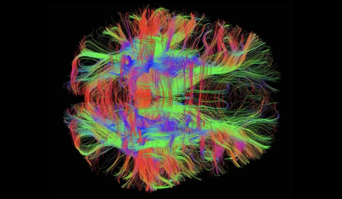
In healthy white matter, water tends to diffuse along nerve fibers (anisotropy), while damage, inflammation, or loss of structure disrupts those patterns. DTI quantifies these changes using mathematical models called tensors, producing tractography images that visually represent neural pathways.
Diffusion Tensor Imaging (DTI) leverages the natural motion of water molecules in brain tissue to map white matter pathways. In organized white matter bundles, fatty myelin sheaths around axons restrict water movement perpendicular to the fibers, allowing freer diffusion along the tract—an anisotropic property that DTI precisely measures.
Key steps in the process include:
Applying magnetic gradients in multiple directions to capture diffusion data from each voxel.
Modeling the data into tensors that represent water movement direction and magnitude.
Generating 2D and 3D tractography maps to visualize neural connections across brain regions.
Two essential metrics quantify brain microstructure: Fractional Anisotropy (FA) measures directional preference of water diffusion, where high values indicate healthy, well-organized fibers and low values suggest damage or degeneration. Mean Diffusivity (MD) captures average water motion, with elevated levels often signaling cellular loss, edema, or tissue breakdown.
DTI reveals details about white matter integrity that standard MRI sequences cannot detect, making it essential for both clinical diagnosis and neuroscience research. By mapping the orientation and health of neural tracts, DTI uncovers subtle disruptions invisible to conventional imaging.
Key applications include:
Traumatic Brain Injury (TBI): Detects microscopic axonal shearing and diffuse injury often missed by routine MRI or CT.
Stroke Evaluation: Tracks white matter damage progression and recovery patterns over time.
Neurodegenerative Disorders: Monitors connectivity loss in multiple sclerosis, Alzheimer’s, and Parkinson’s disease.
Surgical Planning: Visualizes critical tracts to guide safer tumor resection and epilepsy procedures.
Development & Research: Studies brain maturation, autism spectrum disorders, aging, and neural plasticity.
This advanced capability helps clinicians intervene earlier and tailor treatments more precisely.
DTI provides detailed white matter mapping when structural MRI alone cannot fully explain symptoms or guide treatment. Physicians order it to assess microscopic fiber damage and connectivity disruptions across various conditions.
Common applications include:
Traumatic brain injury and concussion diagnosis to detect axonal shearing.
Stroke and ischemic injury assessment to track white matter recovery.
Multiple sclerosis and demyelinating diseases monitoring tract degeneration.
Tumor impact evaluation to see effects on surrounding neural pathways.
Neurodevelopmental disorders and research studies on connectivity.
A DTI scan uses specialized sequences on a standard MRI scanner and follows a straightforward, non-invasive process. No radiation is involved, and comfort levels match conventional MRI exams, typically lasting 45–60 minutes.
Preparation involves removing metal objects and following positioning instructions specific to the brain region being studied. Technologists guide patients through each step to ensure clear images and a relaxed experience. The resulting connectivity maps help pinpoint disruptions and inform precise treatment strategies.
Yes — Diffusion Tensor Imaging is a safe MRI technique that does not use ionizing radiation. The experience is similar to a conventional MRI scan. Most patients tolerate the exam well, and our team is committed to making your visit as comfortable and stress-free as possible.
For car accident victims and trauma patients, DTI can be especially valuable because it helps turn invisible brain damage into visible evidence. Standard imaging may appear normal even when symptoms persist, but DTI can reveal microstructural changes in white matter — aiding clinical diagnosis, treatment planning, and documentation for medical and legal purposes. (Content synthesized from clinical principles and common practice.)
Precision MRI offers advanced Diffusion Tensor Imaging as part of our comprehensive neuroimaging services. With cutting-edge technology and expert radiology interpretation, we help patients and healthcare providers gain deeper insight into complex neurological conditions.
Schedule your DTI scan today to access advanced brain connectivity imaging and take a confident step forward in diagnosis and care.
1. What is Diffusion Tensor Imaging (DTI)?
DTI is a specialized MRI technique that maps the brain’s white matter pathways by measuring the directional movement of water molecules.
2. How is DTI different from a standard MRI?
While standard MRI shows brain structure, DTI reveals the organization and integrity of neural connections.
3. How long does a DTI scan take?
A typical DTI MRI exam takes about 45–60 minutes.
4. Is DTI safe?
Yes — like other MRI scans, DTI is non-invasive and does not use radiation.
5. What conditions may require DTI?
DTI is often used for traumatic brain injury, stroke evaluation, neurological disease monitoring, and surgical planning.