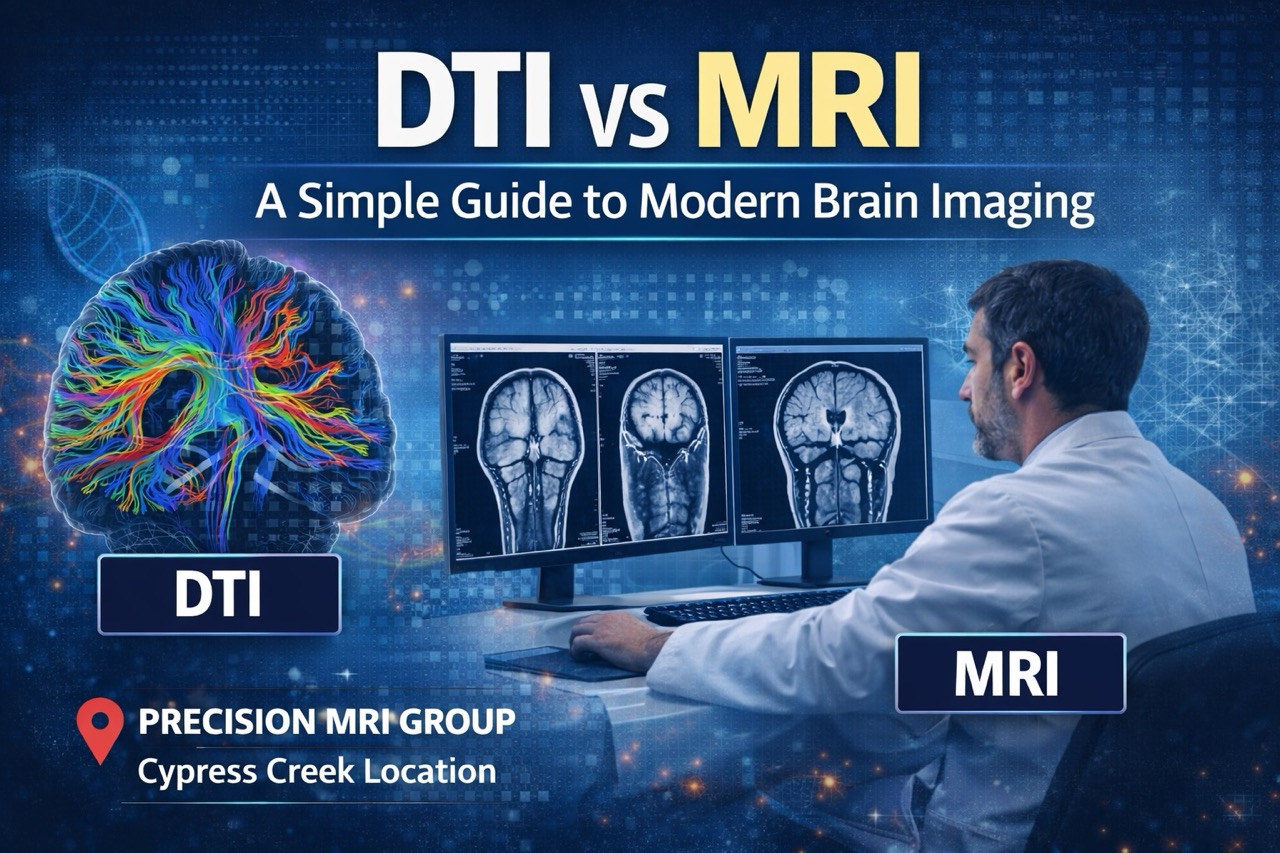
DTI vs MRI A Simple Guide to Modern Brain Imaging
Your doctor ordered brain imaging, but you’re confused about MRI versus DTI scans. Both use magnetic technology, yet they reveal different information about your brain’s health.
Many patients wonder if they need standard MRI or advanced DTI imaging for symptoms. The answer depends on what your doctor needs to see inside your brain clearly. At Precision MRI Group, we offer both imaging types at our Cypress Creek location. Our experienced team helps you understand which scan provides the answers you need most.
What Is a Standard MRI?
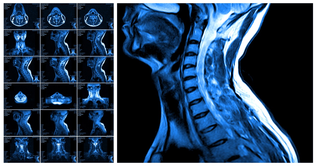
Standard MRI uses powerful magnetic fields and radio waves to create detailed brain pictures. The technology shows brain structure, including tissue, blood vessels, and fluid-filled spaces clearly. Doctors use MRI to detect tumors, bleeding, stroke damage, and structural abnormalities routinely.
Common conditions detected with MRI include brain tumors, multiple sclerosis lesions, and bleeding episodes. The imaging reveals large-scale tissue damage from trauma, infection, or disease processes effectively. Traditional structural imaging excels at showing what brain tissue looks like and identifying obvious problems.
Standard MRI remains the first choice for most brain imaging needs because it’s widely available. Furthermore, MRI provides comprehensive structural information doctors need for many neurological diagnoses successfully.
What Is a DTI Scan?
Diffusion Tensor Imaging, or DTI, is an advanced form of MRI technology for brain evaluation. The scan tracks how water molecules move along white matter pathways connecting brain regions. These pathways contain nerve fibers that carry signals between different brain areas for function.
DTI is considered advanced neuroimaging because it shows microscopic changes standard MRI cannot detect. The technology reveals damage to nerve fiber pathways even when brain structure appears normal. Conditions DTI helps evaluate include mild traumatic brain injury, concussion effects, and white matter diseases.
DTI vs MRI comparison shows that DTI provides functional information about brain connectivity and integrity. Furthermore, this specialized imaging detects subtle injuries that explain persistent symptoms after normal MRI.
How DTI Differs from Standard MRI
Both imaging types use MRI technology but focus on different aspects of brain health. Understanding these differences helps explain why doctors sometimes order both scans for complete evaluation.
Structural Imaging vs Microstructural Imaging
Standard MRI provides structural imaging that shows brain anatomy and obvious tissue abnormalities clearly. DTI offers microstructural imaging revealing damage to individual nerve fibers and pathways invisibly. Structural scans show what brain tissue looks like, while microstructural scans show how it functions. Furthermore, DTI vs MRI distinctions matter most when symptoms persist despite normal structural imaging.
What Each Scan Reveals Inside the Brain
MRI reveals brain tissue appearance, tumors, bleeding, inflammation, and large structural changes affecting anatomy. DTI reveals white matter integrity, nerve pathway disruption, and microscopic damage affecting brain connectivity. Standard scans show problems you can see on pictures, while DTI shows problems affecting how brain regions communicate. Furthermore, combining both provides comprehensive evaluation of structure and function together.
Why DTI Detects Subtle Injuries MRI May Miss
Concussions and mild traumatic brain injuries often damage nerve connections without destroying tissue visibly. Standard MRI appears normal because tissue structure remains intact despite functional pathway damage occurring. DTI detects these connection disruptions by measuring abnormal water movement along damaged nerve fibers. Furthermore, DTI vs MRI comparison shows DTI’s superiority for detecting invisible brain injuries.
When Doctors Recommend MRI, DTI, or Both
Different clinical situations call for different imaging approaches based on suspected conditions and symptoms. Standard MRI works well for acute injuries, tumors, bleeding, and obvious physical brain abnormalities. Post-concussion symptoms and mild TBI often require DTI when standard imaging appears normal.
Stroke-related changes benefit from both MRI showing tissue damage and DTI revealing pathway disruption. Neurodegenerative conditions like Alzheimer’s disease show progressive changes on both imaging types over time. Combined imaging provides a fuller picture when doctors need comprehensive brain health evaluation.
Furthermore, DTI vs MRI decisions depend on symptom type and previous imaging results available.
Advanced DTI Brain Imaging Now Available at Precision MRI Group
Precision MRI Group recently upgraded our Cypress Creek MRI machine to perform advanced DTI scans. This enhancement allows us to offer cutting-edge neuroimaging for patients needing detailed brain evaluation. We added DTI capabilities because many patients have symptoms that standard imaging cannot explain.
The upgrade benefits patients with persistent neurological symptoms after normal MRI results frustratingly. Athletes with concussion symptoms, accident victims with brain injuries, and stroke patients need DTI. Board-certified radiologists specializing in neuroimaging interpret both standard MRI and DTI scans expertly.
Clinical Scenarios Where DTI Provides Superior Insight
Certain medical situations require DTI’s advanced capabilities beyond what standard MRI can reveal. Understanding when DTI vs MRI matters most helps patients and doctors make informed decisions.
Mild Traumatic Brain Injury (When MRI Appears Normal)
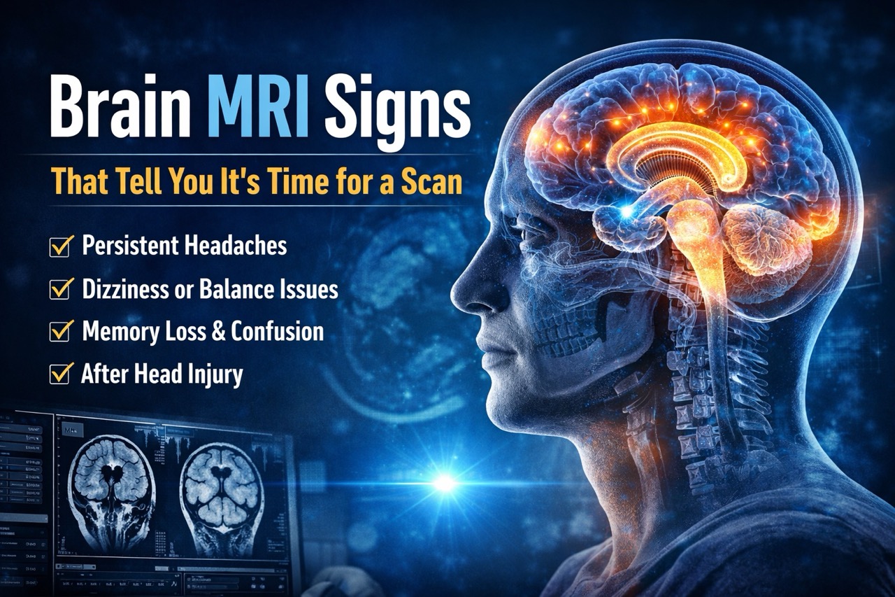
Concussions cause symptoms like headaches, confusion, and memory problems despite normal standard MRI. DTI reveals microscopic damage to nerve pathways that standard imaging cannot detect at all. The scan shows why patients have real symptoms when structural imaging looks completely normal. Furthermore, DTI provides objective evidence of brain injury for treatment and documentation purposes.
Memory Issues, Dizziness, Mood Changes After Injury
Persistent cognitive and emotional symptoms after head trauma warrant DTI evaluation for pathway damage. Standard MRI misses subtle white matter injuries causing these debilitating symptoms in patients. DTI reveals which brain connections are disrupted, explaining specific symptom patterns experienced clearly. Furthermore, this information guides rehabilitation and helps predict recovery timelines more accurately.
Stroke Recovery Mapping and Rehabilitation Planning
DTI shows which neural pathways survived stroke and which were permanently damaged by events. This information helps therapists design rehabilitation programs targeting preserved brain connections effectively still. The imaging predicts recovery potential by showing intact pathways that can compensate for damage. Furthermore, DTI vs MRI comparison shows DTI’s value for personalized stroke recovery planning.
White Matter Disorders and Neural Pathway Disruption
Multiple sclerosis, leukodystrophies, and other white matter diseases benefit from DTI monitoring over time. The scan tracks disease progression and treatment response better than standard MRI alone. DTI reveals early pathway damage before symptoms become severe or irreversible completely. Furthermore, this early detection enables intervention when treatment is most effective for outcomes.
Comparing Image Detail and Diagnostic Value
Standard MRI excels at showing brain anatomy with exceptional detail and clarity for structure. The imaging reveals tumors, bleeding, inflammation, and tissue damage visible to radiologists reviewing pictures. DTI shows connectivity and microstructural integrity of nerve pathways connecting brain regions functionally.
DTI vs MRI comparison reveals that each provides unique, complementary information about brain health. Combining both scans leads to stronger diagnostic accuracy when symptoms are complex or unexplained. Comprehensive evaluation using multiple imaging techniques provides doctors with complete information for treatment. Furthermore, advanced imaging prevents misdiagnosis and ensures appropriate care based on actual conditions present.
What Patients Can Expect From a DTI or MRI at Precision MRI Group
Both standard MRI and DTI scans at our facilities provide comfortable, patient-focused experiences. The procedures are painless and follow similar protocols for positioning and image acquisition. Late-night and weekend scheduling accommodates busy work schedules and urgent evaluation needs without delay.
Free transportation upon request removes barriers for patients who have difficulty getting to appointments. Multilingual staff speaking English, Spanish, and Creole ensures clear communication with all patients. Reports are delivered within 24 to 48 hours, with immediate results available upon request. Furthermore, our Cypress Creek facility offers both MRI and DTI for comprehensive neurological evaluation.
Get the Brain Imaging You Need at Precision MRI Group
Accurate diagnosis requires choosing the right imaging test for your specific symptoms and situation. Advanced imaging like DTI becomes essential when standard tests leave questions unanswered about health. Precision MRI Group leads South Florida in neuroimaging technology with both standard and advanced capabilities.
Our commitment to offering cutting-edge diagnostic tools ensures you get answers when symptoms persist. Don’t settle for incomplete evaluation when advanced imaging can reveal what’s really wrong.
Schedule your MRI or DTI scan at Precision MRI Group and get clear, dependable imaging results from a caring team dedicated to your health.
Precision MRI Group Locations:
Pembroke Pines
9696 Pines Blvd., Pembroke Pines, FL 33024
Phone: (954) 391-7844, Contact: Amalia (amalia@pinesimagingcenter.com)
Lake Worth
2311 10th Ave N Suite #2 and Suite #1, Lake Worth, FL 33461
Phone: (561) 623-8346, Contact: Marisol (marisol@mriprecision.com)
Cypress Creek
2122 NW 62nd Street, Suite 107, Ft. Lauderdale, FL 33309
Phone: (954) 677-1069, Contact: Latoya Reid (latoya@cypresscreekmri.com)
Port St Lucie
879 E Prima Vista Blvd #2, Port St. Lucie, FL 34952
Phone: (772) 344-7566, Contact: Laura Schwenzer (laura@mriprecision.com)